
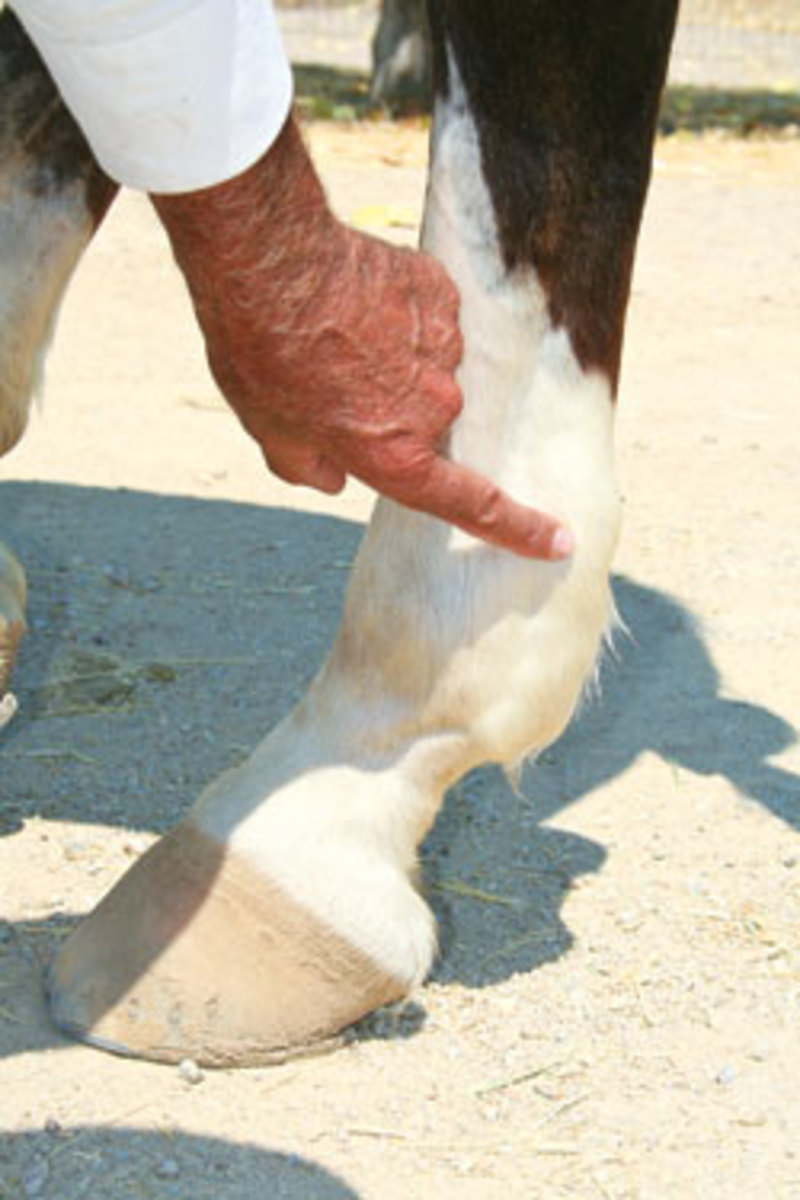
The digital tendon sheath is a structure that surrounds the flexor tendons as they course behind the fetlock. The sheath begins approximately four inches above the fetlocks, and extends distally to the lower pastern. The sheath has a synovial lining similar to a joint, and allows the tendons to slide freely during extension and flexion of the fetlock.
In my observation, many horses can develop a distention of this structure that is benign, in that it never causes a lameness problem. However, I have seen horses develop career-ending problems when progressive changes build up to cause degenerative problems in the deep flexor tendon. To explain the difference between a cosmetic blemish and a functional unsoundness, I will attempt to describe the pathophysiology of the problem.
Repetitive stress and strain can cause the cells that line the internal wall of the sheath to produce abnormal amounts and type of synovial fluid that stretches or distends the sheath. In progressive cases, micro-hemorrhage or extravasation of protein-rich fluid can lead to adhesions (a form of scarring) between the wall of the sheath and the peritendinum (covering) of the deep flexor tendon. With this situation, you can have impaired motion during athletic activity and continually cause more inflammation and scarring, which ultimately leads to degenerative problems of the flexor tendons.
Identifying when you have the threatening form of this injury can be problematical. Distention of the structure is the first sign one usually notices with the problem. A combination of rest, anti-inflammatory medication and supportive bandaging to allow healing is usually adequate treatment at this stage.
In cases where flare-ups occur or any sign of lameness relative to this condition is evident, more invasive treatments are indicated. Draining the excess fluid and injecting the sheath with agents used in joint inflammation are done. In the most severe cases, surgical intervention is indicated. The surgery may be in the form of arthroscopically cleaning up the adhesions between the sheath wall and tendon, as well as relieving pressure on the swollen structures by resecting a ligament in the area. Because of the range of threats from this condition, evaluation and monitoring by your veterinarian is certainly indicated.


