Tendons of muscles in horses that cross high-motion joints have synovial sheaths around them to facilitate the movement of the tendons up and down with extension and flexion of the limbs. The tendon sheaths in horses I would like to concentrate on in this column are the ones around the flexor tendons in the area of the fetlocks, both front and rear.
The flexor tendon sheathsin horses in the area of the fetlock are the ones that present the most threat to soundness in the horse. Trauma to these structures, either in the form of a strain or internal tear or from a penetrating wound, can lead to serious consequences.
The inside of the sheath is lined with cells that secrete synovial fluid, similar to the lining of a joint, that help lubricate the structure. The lining is also rich with sensory nerve endings that cause significant, sometimes incapacitating, pain when inflamed. When traumatized significantly, you can start an inflammatory process that results in adhesions or scar tissue between the wall of sheath and the tendon it covers. This situation can result in a chronic, painful condition that is difficult to manage in an athletic horse.
The internal type of injury results from a strain that causes some bleeding or leaking of fibrin into the space of the sheath. These products are the precursors to the formation of scar tissue that causes adhesions. Once you have adhesions in this structure, you can start a cycle of repeated tearing and more scar tissue forming because the adhesions are not elastic and are therefore subject to stress trauma with athletic use.
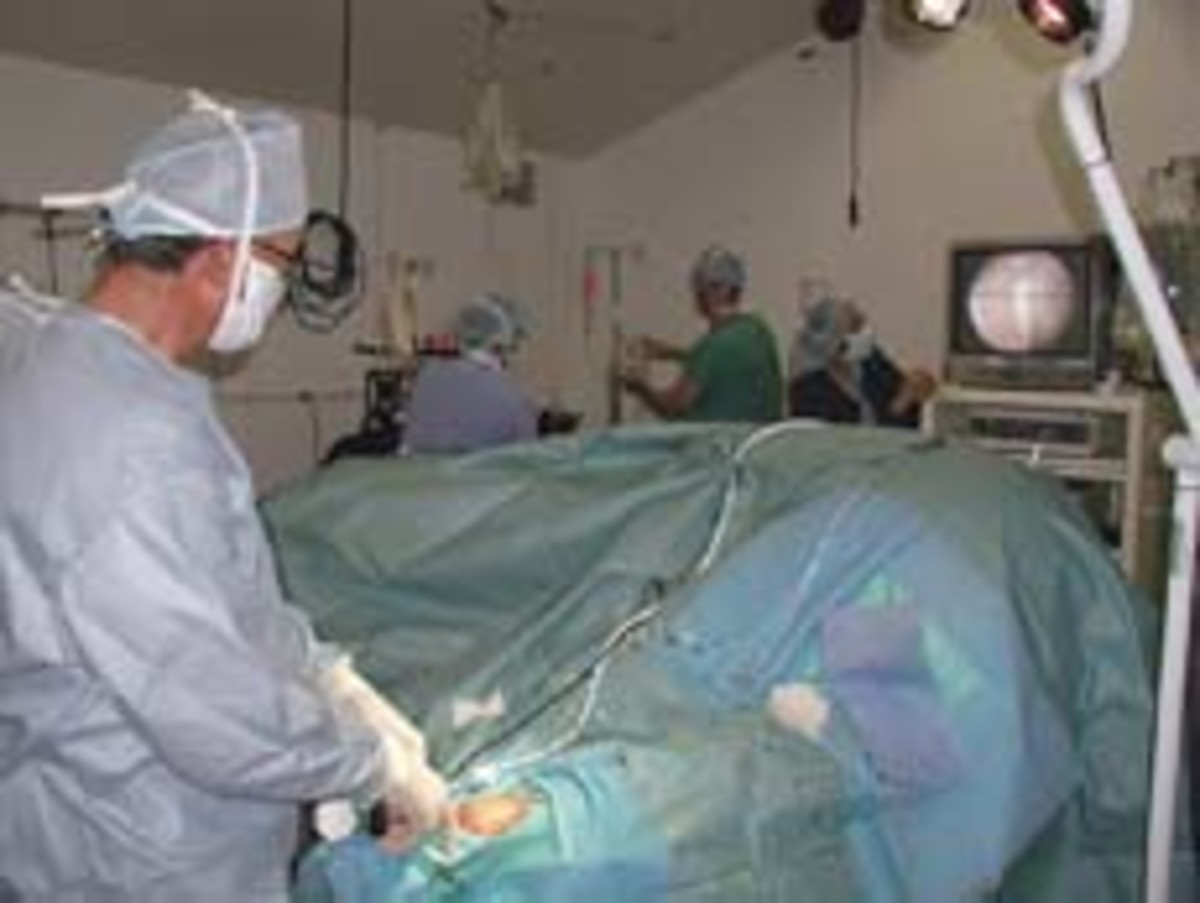
The goal in treating this problem is to minimize scarring and adhesion formation as early as possible. The success is based on early detection and doing things to prevent scarring. These things would include rest, anti-inflammatory medications (both local and systemic), and supportive bandaging to help reabsorb the products of inflammation before permanent changes take place. If you have already reached the point of having severe adhesions, there is the alternative of arthroscopic surgical removal of the adhesions.
The threat of having this condition evolve from a penetrating wound is also significant. Sometimes a small puncture wound that introduces infection into this structure can be disastrous. The flexor tendon sheath starts two to three inches above the fetlock and goes down to the mid-pastern area. If you notice a wound in this area, I advise serious and immediate intervention.
I routinely put horses on systemic antibiotics and anti-inflammatory drugs as well as local debridement and bandaging over an antibiotic dressing. I don’t usually attempt to suture these wounds because, due to their location, they’re usually already contaminated with bacteria when noticed. If you close these contaminated wounds, they often blow up with significant infection.
I think it is important to use enough pain medication to have the horse putting full weight on the affected leg to prevent adhesions forming with the ankle in a “cocked” or flexed position. Having weight-bearing on the affected leg also protects the opposite leg from potential disastrous effects of having all the weight constantly. Interestingly, I’ve noticed this problem more often in the hind legs than the front.
For more articles by Frank Santos, DVM visit: http://www.teamroper.com/Education/Default.asp?Action=View&SubAction=EdArticle&ID=88


