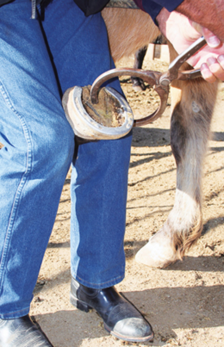
“Navicular disease” is a very common and dreaded syndrome in the horse. It may be the most commonly diagnosed lameness in Western performance horses. The diagnosis and treatment of this problem has been controversial historically.
The diagnosis of navicular disease has traditionally been based on clinical observation, localizing the source of pain with diagnostic nerve blocks, and radiograph (X-ray) interpretation of the navicular bone. Through advanced imaging techniques, mainly MRI, the soft tissue structures in the navicular area have been shown to play a significant role in this syndrome.
There often are other sources of pain causing lameness in this area than just an affected navicular bone. In a recent column, I said colic was not a specific diagnosis, but just a term describing abdominal pain. I think we (veterinarians and horse owners) often use the singular term “navicular disease” for what is really a complex problem. The realization of the complexity of the problem seems to me to explain the differences we often encounter in the response to treatment. If there can be multiple causes for the symptoms, how can there be any one way to treat it?
Another complicating factor with this problem has been interpreting what is on radiographs of the navicular bone. There are natural variations in the navicular bone of apparently normal horses. There are some horses with questionable changes radiographically in the navicular bone that are perfectly sound.
Then, there are horses with normal looking radiographs of the navicular bone that are chronically lame from pain emanating from the navicular area. With techniques that can image soft tissue structures in this area, it becomes evident that a large number of affected horses have lesions in these soft tissue structures that are causing the problem.
Structures such as the deep flexor tendon, the bursa between the deep flexor tendon and navicular bone, and the ligaments supporting the navicular bone can be affected with or without changes in the navicular bone itself.
This information shouldn’t overwhelm or confuse, but should add to the understanding of the complexity of this problem. I think that as more diagnostic aids in imaging these structures become available, it will be possible to define what the specific lesion is, and therefore be better able to treat it.
For instance, a horse that incurs an injury to the deep flexor tendon in this area may benefit from time and rest to allow healing. Often, horses may benefit from local injection of a therapeutic agent for a bursitis problem.
I don’t want to imply here that there will be a cure for every horse. But more informed decisions can be made regarding treatment and prognosis.
Finally, I hope this information helps dispel the myth that radiographs alone tell the story of the status of “navicular disease.” SWR