
Historically, horses with chronic or recurring heel pain were often diagnosed as having “navicular disease.” I put the term “navicular disease” in quotes for a reason. People came to think of this syndrome as a single entity. With advances in new modalities of imaging the structures in this area of a horse’s foot, it is evident that there are many different lesions or scenarios that may be causing the problem. Therefore, I believe it to be an oversimplification to say a horse has “navicular disease,” per se.
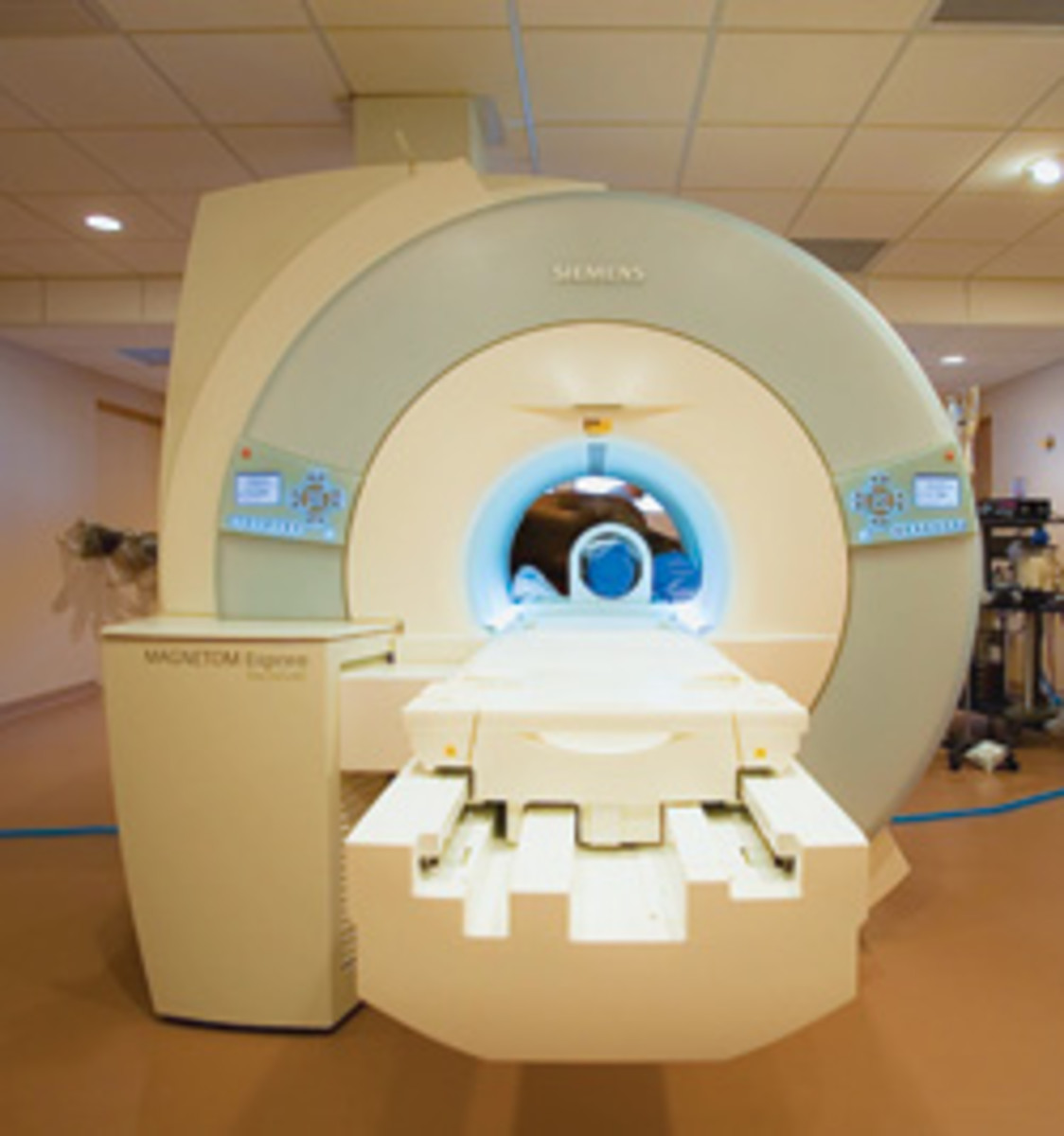
Having the ability to identify what specific lesion is causing the problem is a necessity to make a logical decision as to treatment and/or prognosis for the future. I recently consulted an orthopedic surgeon regarding a shoulder problem. His clinical examination and radiographs (X-rays) of my shoulder weren’t satisfactory, in his opinion, for an absolute diagnosis. So he referred me to an imaging center for an MRI appraisal of my shoulder. I believe, in the future, this scenario will be more commonly used with horse problems. However, presently there are very few MRI units available for use in equine diagnostics.
An MRI evaluation of a horse’s foot not only shows the status of bone structures, but also the so-called “soft tissue” structures, such as the deep flexor tendons and various ligaments in the area. These structures are often shown to be the cause of a problem, and explain why some horses are chronically lame with heel pain but have “normal” radiographs. As I mentioned earlier, the availability of MRI diagnostics in horses is limited because of the cost factor. The horse also has to be anesthetized for the procedure, to be still for the time required, and only the head and distal limbs of a horse fit into our present machines.
Another mode of soft tissue diagnostics that is evolving for lameness problems is ultrasonography. The first use of the ultrasound machine in horses came about 25 years ago for diagnosing early pregnancy. With advances in technology and experience, many structures such as tendons, ligaments and intra-articular defects can be evaluated with this modality. While these machines aren’t cheap, they’re much more affordable than the MRI unit. Ultrasound machines are also portable, and it’s a non-invasive procedure.
I firmly believe these modalities of diagnostics are going to make more exact diagnosis of heretofore vague problems possible. The cost will not be small, due to investment in the machines and the training needed to properly use them. But veterinary medicine will advance because of it.


